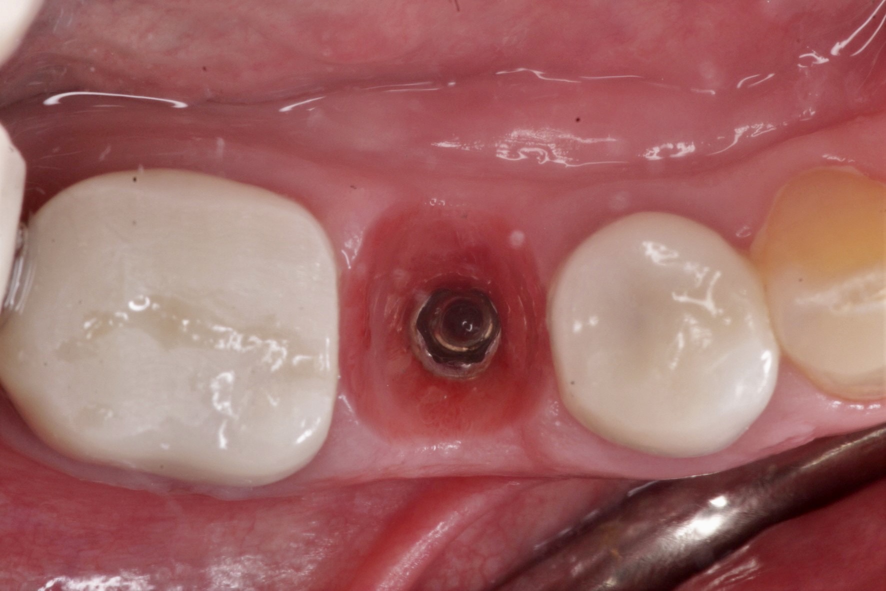
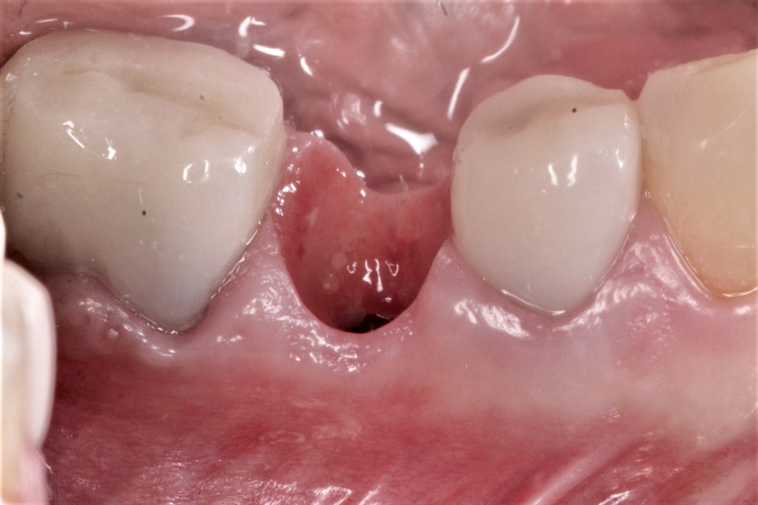
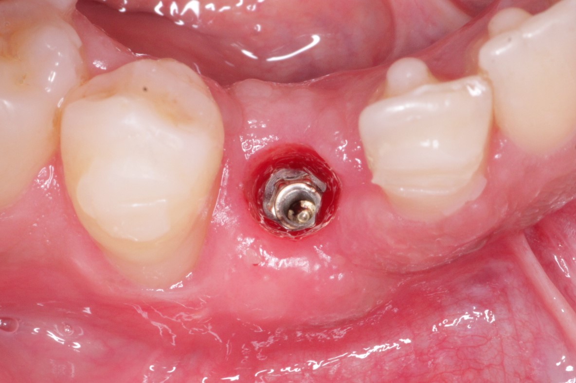
Studies have shown that there are many factors that determine the tissue contour and papilla fill between implants and teeth. But it can be argued that the best way to do so is to preserve the tissue contour from the very beginning. In this case, an immediate implant was placed into the socket followed by the fabrication of an immediate screw-retained provisional. 3 months later, the provisional was removed so that an impression could be taken and the peri-implant tissue was almost an exact replication of the original socket (1st two photos below). This can be difficult to achieve when a standard healing abutment is placed instead (last photo). What techniques and recommendations do you have for maximizing the chances of natural peri-implant tissue?
I completely agree. But why would you immediately load instead of placing a healing abutment shaped with composite added to fill the socket? Unless you feel that the risks of the patient not following the recommendations would have the same consequences whether it’s a temporary crown or a healing abutment!?
Btw what are your recommendations in a case like this?
Waiting for your response.
Hi. Great question. If there is no esthetic demand, then your suggestion of an custom anatomic shaped healing abutment would work just as well without the increased risk of immediate provisionalization. For single units like this, the provisional is always out of occlusion to avoid “loading”.
And what are your instructions regarding what not to eat and for how long!?
Sorry to say but I think you had used healing abutment with small width. You should use large width abutment it would give the same result as you achieve with provisional
@S_Revach as long as the provisional is out of occlusion in centric/excursive/protrusive then its just a matter of making sure the patient is aware of it and favors the other side of the mouth as much as possible when chewing.
@jamshid_khan No reason to apologize for your opinion. A wider healing abutment in this case may give you more emergence on a horizontal plane but it would not preserve the vertical dimension of the papillas and buccal and lingual tissue scallop.