Hello dear colleagues, your opinion is needed.
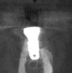
Healthy elder female presented with a case of mandibular atrophy in the posterior region. Patient wanted only one implant to be inserted to have an antagonist with the opposing teeth for better masticatory function. An attempt was made to insert a shorter implant (4x7mm). Osteotomy was performed using Versah drills, implant was placed several mm subcrestally with good primary stability (torque wasn’t too high, aprox 35Ncm). Since the buccal wall was very thin but intact, an attempt was made to decompress soft tissues using membrane, buccal bone was grafted with allograft. Primary closure was achieved. Following up no problems were observed around the wound. 6 months later implant was uncovered, healing abutment was placed, however, upon closer look on buccal wall exposed implant threads were observed. On the CBCT scan buccal bone defect is visible, around 1/2 of the implant length. Implant is osseointegerated.
Now I understand that proper GBR should have been done, maybe other approaches should have been used, trust me, me of all people now clearly knows this. My question is - what’s next? Buccal threads are not showing through gingiva (yet, but I expect them to show in a year if not faster), most of the implant is in the bone, however I’m very concerned over functionality of this implant. CTG graft and continue with restoration? Perform additional GBR, if so, what bone substitute would you recommend? All opinions and criticism are very welcome, I kindly ask your general opinion on this matter. Thank you.
PS sorry for not including clinical photos of the case, I may be able to take them before and after recommended treatment, unless of course general opinion will be to remove the implant
You don’t need to do anything as long as the surrounding soft tissue is adequate. While this may not have been your desired outcome it is actually quite normal and should pose no problems in the presence of adequate soft tissue attachment. Leave it alone and stop taking post treatment CBCT because often times that extra grafting that is done around an otherwise stable newly inserted fixture will show itself just like this… Think back to the nature of the area that you grafted during the initial placement. That very thin but intact buccal wall that you sprinkled boe dust on was avascular cortical bone which had been further compromised by the compression of the Versah protocol. The graft never had a chance but it was not necessary in the first place… as long as the soft tissue is adequate. It is fine!!!
I would be more concerned about your protocol " implant was placed several mm subcrestally with good primary stability" as this unnecessary subcrestal placement appears to have put you dangerously close to the IAN…
Thanks for a quick response! I have no idea whether it is lack of my own experience with the system, but I get very varying results using osseodensification. Some turned out acceptable, some disastrous, especially in posterior mandible. As far as I remember this was last case I did using that protocol, likely it will be the very last case. Even with maxillary cases I use other systems for closed sinus lifts and they turn out to be more predictable in my opinion at least.
Regarding this case, I will make sure to improve soft tissues if need arises, thanks for your opinion. Looking forward to hearing more thoughts on this case from other colleagues
And I agree with you 100 percent, implant was positioned very close to the nerve. I usually never operate this close to IAN. I’ve seen other cases where canal is much further away from the implant and some neurological symptoms appear.
This could be my own lack of experience as I have never bought into the whole osseodensification concept but I can’t imagine it having any value in the mandible any more than aggressive threaded fixtures in the mandible. While it doesn’t fit the current trend which places entirely too much emphasis on primary insertion torque the most successful implant fixture of all time is a parallel walled tissue level Straumann/ITI design. I say this as someone who does not routinely utilize parallel walled tissue level fixtures, simply because my referrals are not familiar with them. When we ignore the fact that most of these old school successful fixtures are placed <30Ncm and still survive despite also having a one stage component built into the system it makes me wonder why we go through so much trouble as to incorporate Versah in the first place.
As long as the implant is stable. Best is not to touch it. The more you open it. the more bone loss will happen. hopefully the implant will out live your patient. Next time just use a smaller diameter implant. implant diameter is determined by alveolar ridge thickness. This patient’s cortical plate is too thick to use versah burs to try to expand it. this is probably why your cortical bone become necrotic and lost it.
This case actually demonstrates a major flaw in the Zero Bone Loss Concept as it is promoted by the implant manufacturers. This is a posterior tooth and there is no reason, other than the protocol, to have placed this fixture subcrestal. Because the concept is widely promoted yet grossly misunderstood this is an unfortunate, not catastrophic, and common problem when blindly following the protocol. This fixture, by virtue of following a cookbook protocol, was unnecessarily placed entirely too close to the IAN and an excess amount of the buccal plate was compromised and lost. Both of these could have been minimized by placing the fixture crestal and allowing the natural platform switch/medialized abutment built into the system to actually do its job which has been demonstrated to adequately preserve crestal bone. Nothing wrong with the concept but maybe some things aren’t broke bad enough to really need fixing…
Scotty
keeping up our other discussion about Crestal/Sub-crestal placement, you are 100% correct in this case. If the Cortical plate was not Expanded, it Could have worked. We can spaculate that during Expasion, there was possibly fracturing of buccal plate and it led to this bone loss since Cortical bone has minimal blood supply and bone graft drifted or was resorbed.
So, I guess the take away, the learning point is If you have type 4 type of bone and thick Cortical plate, on CBCT, plan on a Crestal placement, so as NOT to follow a Formula for implant placement. Agreed.
With the Caveat, Hindsight is 20/20, but starting now we Should take your point about crestal/sub crestal placement depending upon type of bone.
I think another important point about following a cookbook placement protocol is that it must be modified when working around structures such as the IAN. Because this was placed using the Neodent’ish subcrestal protocol then the fixture, by virtue of the recipe, had to be 2mm shorter than what the patient’s anatomy would otherwise allow. Now the resulting buccal bone loss, compounded by the osseodensification, is happening to a shorter implant resulting in a greater percentage of non-integrated surface area.
Albatros2: I wonder 1) did you do corticotomy on the buccal to get bleeding from the endosteum?
2) did you tac the membrane down so there is no micro motion?
3) clean the surface of the bone with a acrylic burr to get all the soft tissue tags removed before bone grafting?
First I would want to be sure with either choice that you have adequately thick, soft tissue (1.7-2mm) which you’ve stated isn’t thick enough and adequate height of keratinized tissue (2-3mm). Since you believe the choice of watching it will result in extremely thin tissue, it isn’t a stable soft/hard tissue complex. With what you’ve said, there isn’t enough volume of soft and hard tissue, hence enough vascularity to replace the lost PDL, over the implant to stand the test of use and time. You could correct the defect using GBR noted above and do a simultaneous CTG if there is enough KG. IF there isn’t enough KG, then do the GBR, wait 2 months and do a FGG.
With that much bone missing, I’ll predict that over the next 5 years you’ll see recession/mucositis develop if left “as is” and the rough implant surface will have bacterial contamination. For me, treating it now is more predictable, less painful and an easier surgery. Once any form of peri-implantitis enters into the mix, then getting the bacteria cleaned out before GBR/CTG is a chore. Good luck.
Also once implant body is exposed to oral cavity. meaning not inside bone. you will have biofilm covering on the exposed implant surface and GBR will not work at all.