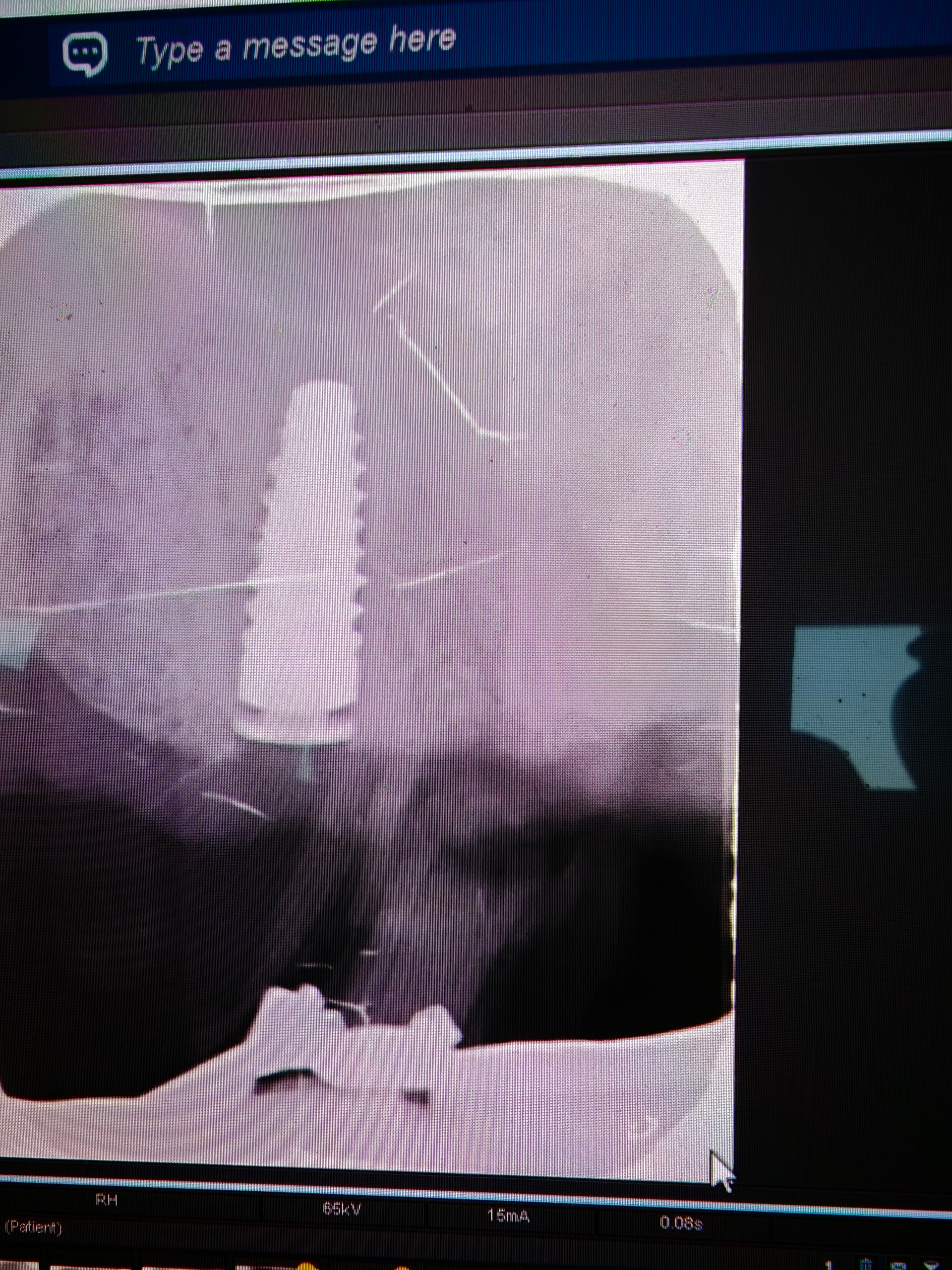
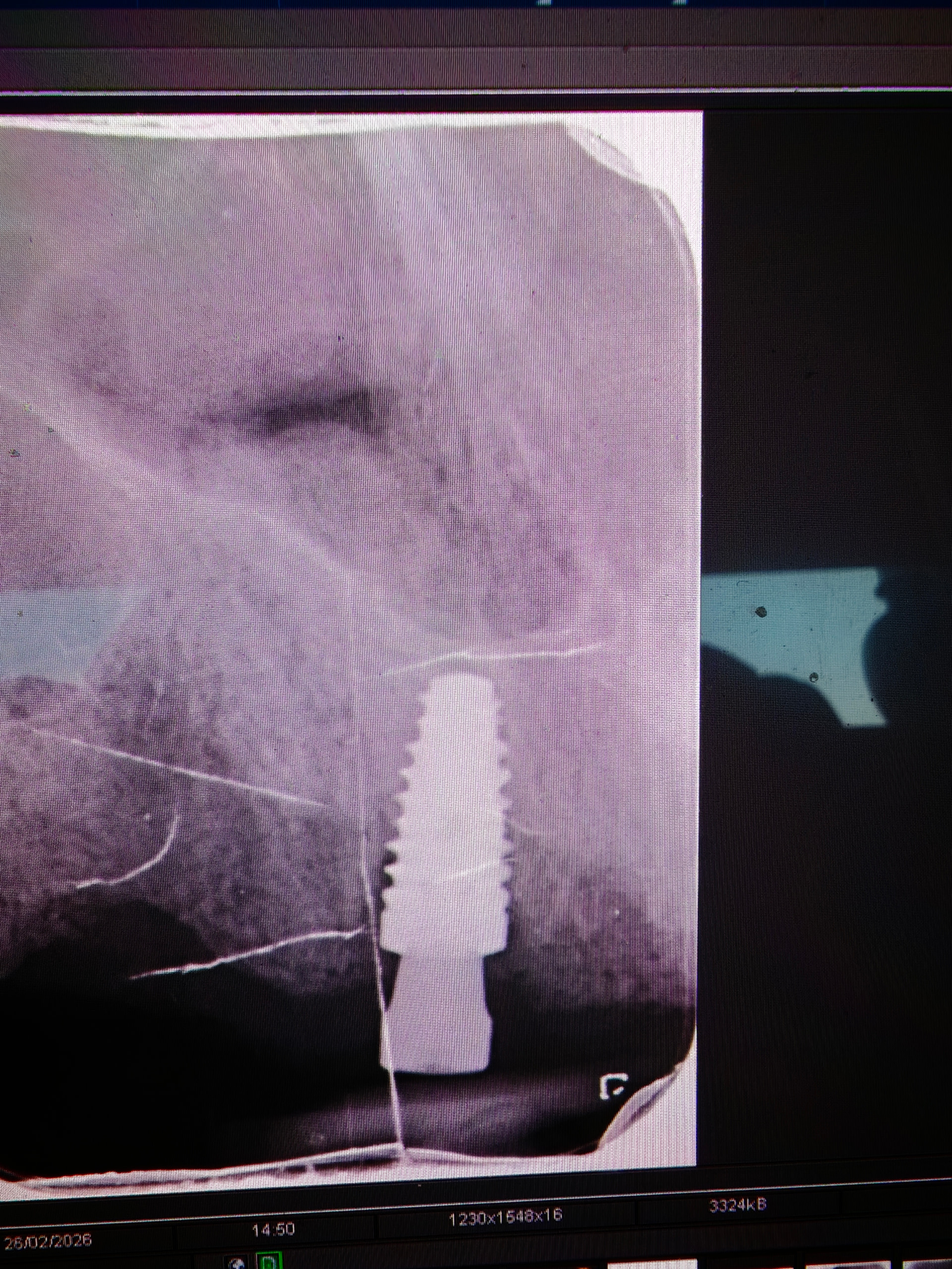
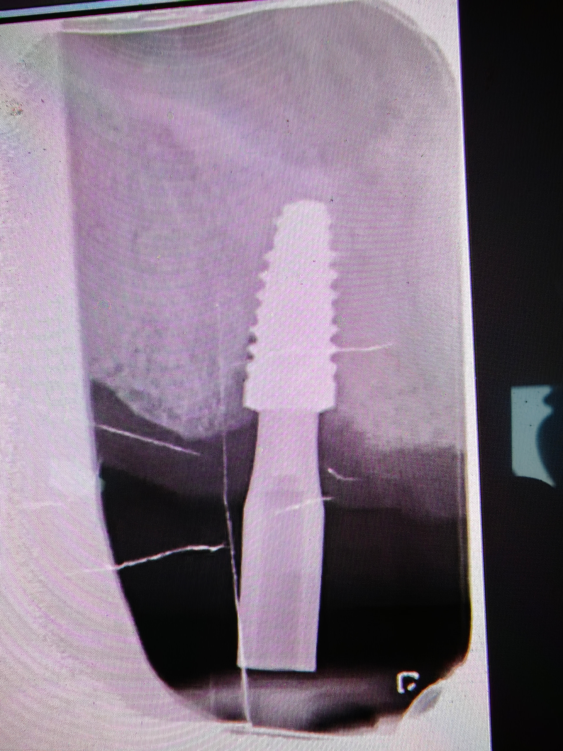
How do I fix this botch job ? There was already a failed implant already near this site therefore switched from BLT to BLC. Initial photo time of placement cover screw not seated which was subsequently fully at the time. Thentime. Then 3 months later with (scan body photo) clearly some massive bone resorption. Restorative dentist says was fine at the time of healing abutment removal and scan body. Patient returned saying healing abutment loose when colleague tightened pain deep inside palate (final photo with healing abutment). Restorative not sure if the implant moved at the time of replacing healing abutment. Is there anyway I can salvage this ?
What does it mean that you are not sure if it moved? Either it moved and then the implant must be removed, or it did not move and you can proceed with prosthetic treatment or solve bone resorption.
I wasn’t the one who assessed the general dentist informed me of that. Anyway I assessed it today it was stable obviously they amount of bone loss so early is of concern or not ?
So the implant was stable and no bleeding on probing but the general dentist decided to send for a CBCT scan and he is concerned to restore since the implant is placed for an implant over denture.
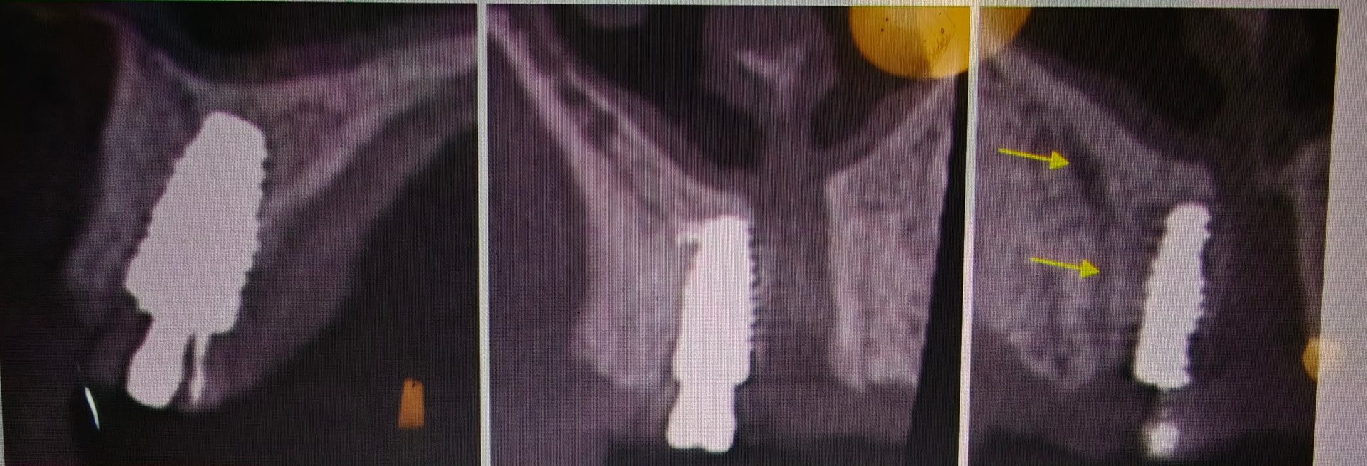
Unfortunately not ideal placement or there’s been a loss of palatal bone. Patient was asymptomatic and as mentioned implant rigid, Should I re enter and graft or leave and continue to restore and we monitor it ? Any advice would be much appreciated
Unfortunately taking 2D images from different angles can/will show vastly different results…The scan body image looks much better than the healing abutment image and I can only conclude that is because it is a fixture placed in soft posterior maxilla bone with images taken from different angles. I think that “We” as trained dental professionals should be more concerned with treating the patient rather than the patient’s x-ray… especially patient’s non standardized x-rays… As far as the CBCT that you just posted I would suggest that you get used to being upset about the degree of palatal/buccal bone shown on a CBCT in the posterior maxilla because it is a “horrible” method to evaluate a fixture placed in this area and will “always” underestimate. On the flip side a CBCT will also frequently justify the unnecessary additional grafting procedure which is a big selling point when it comes down to the decision of ROI. After over 20 years of placing and restoring implants I am more convinced than ever that the tap test, tapping a mirror handle on the healing abutment, with a resulting high pitch ping is still the most diagnostic and accurate method we have to determine successful integration.
Hi scotty I’ve uploaded some screenshots of the CbCt please be gentle.
Always informative and insightful thank you